
New neuroscience method disentangles crossing fibers in brain tissue
02 April 2020
To understand the structure and function of the brain, we need to study the highly complex, three-dimensional pathways of nerve fibers. For a correct reconstruction of the fiber pathways, it is crucial to know the details of fiber architecture at microscopical level, especially in regions with crossing fibers.
One of the most powerful methods to reveal the three-dimensional fiber architecture is 3D Polarized Light Imaging (3D-PLI), a neuroimaging technique developed at Forschungszentrum Jülich, Germany. The fiber orientations are reconstructed by shining polarized light through hair-thin brain sections and measuring the resulting intensity changes. 3D-PLI is used, for instance, in the European Human Brain Project to investigate the 3D fiber structures of the brain in unprecedented detail. But even with this method, scientists so far struggled to interpret the data when it came to regions with crossing nerve fibers.
Now, Miriam Menzel and colleagues at Forschungszentrum Jülich have shown that a relatively simple application of optical light scattering can be used to determine crossing nerve fibers and even fiber crossing angles, providing a major enhancement for the reconstruction of complex nerve fiber architectures in the brain. Besides transmission microscopy measurements of various brain sections, the scientists used biophysical modeling and simulations on Jülich supercomputers to explain their experimental observations and develop new imaging methods.
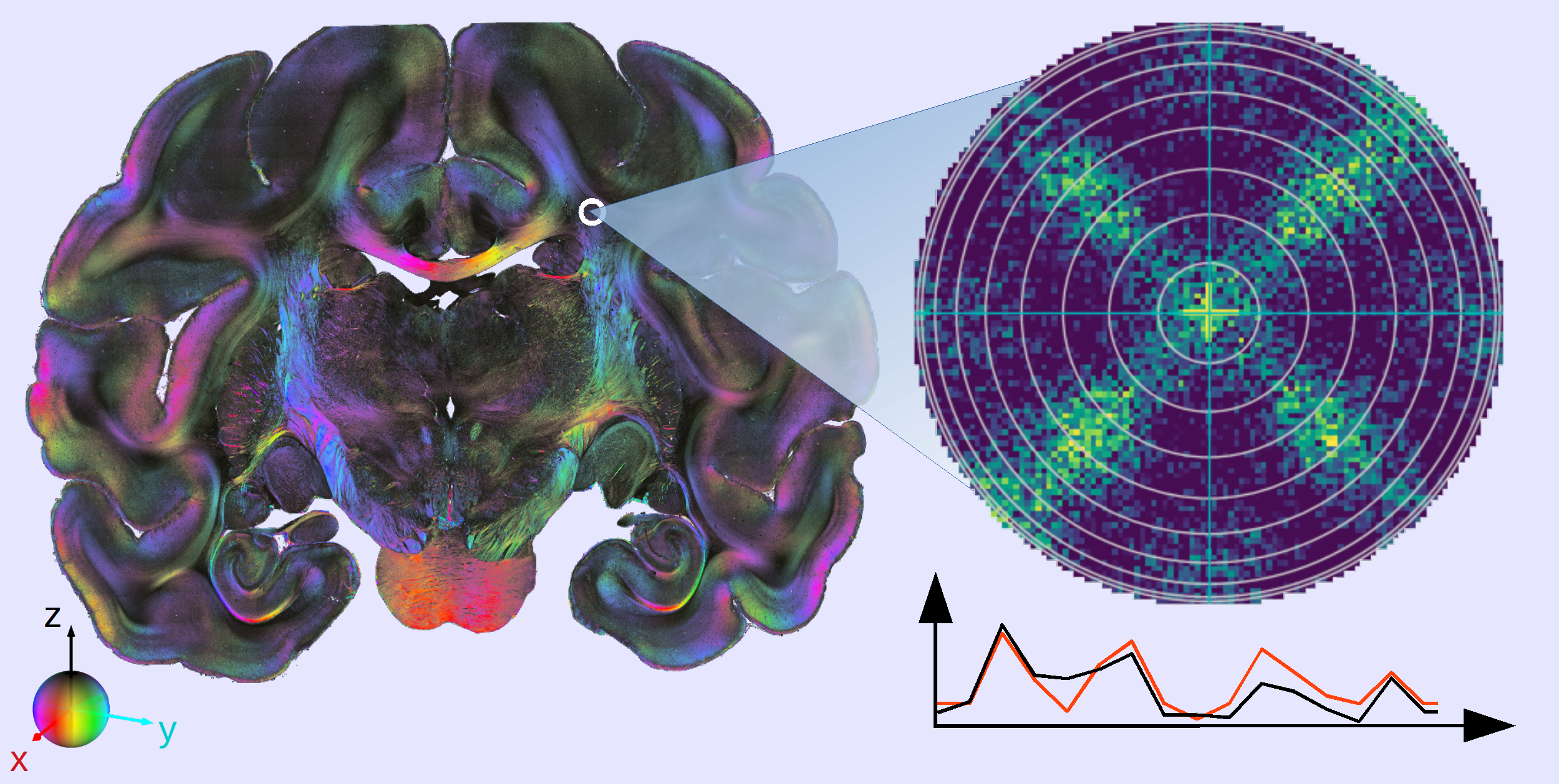
3D-PLI measurement, here pictured on the left in a vervet monkey brain section, reveals the three-dimensional pathways of nerve fibers with microscopic resolution. In combination with light scattering measurements, scientists can now even resolve crossing fibers (right).
Copyright: Forschungszentrum Jülich / Miriam Menzel
Menzel and colleagues found that the intensity of light transmitted through the brain sections depends strongly on the angle at which the light shines on the nerve fibers, but barely on the crossing angles between the fibers themselves. The discovery allows scientists to re-analyze their data sets without repeating any measurements.
Furthermore, the researchers showed that dedicated light scattering measurements can reveal even more details at microscopic resolution like the crossing angles of the nerve fibers– leading to new insights and a deeper understanding of brain connectivity.
Katrin Amunts, a co-author of this work and scientific director of the Human Brain Project emphasizes that advances in methods addressing brain connectivity are key to understand its complex fiber architecture in health and disease.
Part of the HBP Human Brain Atlas
The high-quality data contributes to the multimodal, multiscale Atlas of the Human Brain Project. Neuroscientists and supercomputing experts work together for this to handle the extremely large datasets. The resulting data and analytics tools are available to researchers around the world via the HBP’s digital EBRAINS infrastructure for neuroscience.
Original publication:
Menzel M, Axer M, De Raedt H, Costantini I, Silvestri L, Pavone F S, Amunts K, Michielsen K. Toward a High-Resolution Reconstruction of 3D Nerve Fiber Architectures and Crossings in the Brain Using Light Scattering Measurements and Finite-Difference Time-Domain Simulations. Physical Review X 10, 021002. Published 2 April 2020.
DOI: https:/www.doi.org/10.1103/PhysRevX.10.021002
Further information:
Research group "Fiber Architecture" (FA), Institute of Neuroscience and Medicine, Structural and functional organisation of the brain (INM-1)
Other news:
German-Canadian collaboration boosts the HBP Brain Atlas
MRI and AI to develop a brain virtual biopsy tool
New imaging technique provides structural information about brain tissue
Contact:
Dr. Miriam Menzel
Institute of Neuroscience and Medicine, Structural and functional organisation of the brain (INM-1) Tel.: +49 (0)2461 61-96359
E-Mail: m.menzel@fz-juelich.de
Press Contact:
Peter Zekert
Human Brain Project
Public Relations Officer
Email: press@humanbrainproject.eu