EBRAINS-powered study reveals unknown complexity in avian brains
24 September 2020
An innovative microscopy method shows surprising similarities between mammals and birds
Some birds can achieve extraordinary cognitive performance – but their brains were considered to be rather disorganized compared to those of mammals. Scientists from Bochum (RUB), Düsseldorf (HHU), Jülich (FZJ), and Aachen (RWTH), now for the first time, show striking similarities between the neocortex of mammals and sensory brain areas of birds: Both are wired in horizontal layers and vertical columns. The finding refutes 150-year-old assumptions. Decisive insights were provided by a method developed by Jülich and Düsseldorf brain researchers. The results have been published this week in the journal "Science".
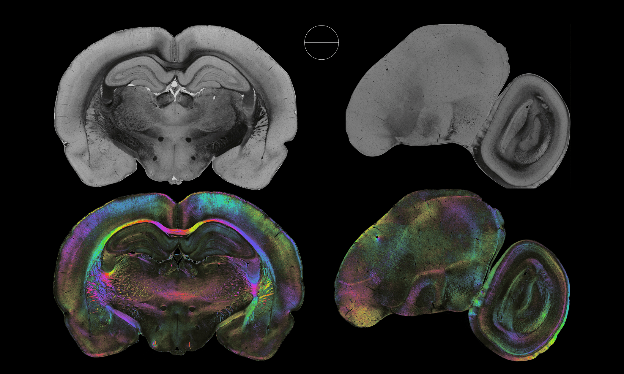
Nerve fibres in the brain of a rat (left) and a pigeon imaged with 3D Polarized Light Imaging Copyright: Axer et al., Forschungszentrum Jülich
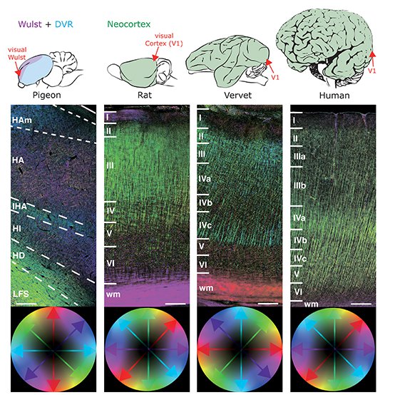
Birds and mammals have the largest brains relative to their body size. Otherwise they have little in common, so the assumption of scientists for more than hundred years: Mammalian brains have a cerebral cortex that is made up of six horizontal layers and columns that run perpendicular to these layers. The avian brain in contrast appears to be poorly organized at first glance, and only shows accumulations of cells with more or less density.
"In view of the astonishing cognitive performance that birds can achieve, however, it was suspected that their brain is higher organized than previously assumed," says Prof. Dr. Onur Güntürkün, head of the Biopsychology work unit at the Faculty of Psychology at the RUB and an expert on the cognition of birds.
Indeed, researchers around Dr. Christina Herold (C. and O. Vogt Institute for Brain Research, HHU) and Dr. Martin Stacho (RUB) have now succeeded in proving that the brains of birds and mammals look surprisingly similar in their organization. The fibers in the bird brain run horizontally and vertically, just like in the neocortex of mammals.
The fiber structure of pigeons and various mammals in comparison. The 3D-PLI method shows the directions of the nerve fibers color-coded. The depictions seen here do not reflect the true sizes - a human brain is about 500 times larger than pigeon brain.
Copyright: Herold et al., HHU Düsseldorf
Previously, it was not possible to map the fiber structure of larger areas of the avian brain with the required accuracy. Commonly used techniques are either limited to small tissue samples or lack resolution and sensitivity to reveal microstructural features that define the brain’s neuronal organization. The level in between therefore remained in the dark.
"3D PLI has a slightly lower resolution than tracing methods, but is capable of analyzing large tissue volumes in a reasonable time – a decisive advantage," explains Dr. Markus Axer, head of the fibre architecture group at the Forschungszentrum Jülich. Using this method, the researchers were able to analyze three complete pigeon brains at a resolution of 1.3 micrometers (millionths of a millimeter). Per brain, 250 sections were scanned at high resolution and reconstructed in 3D.
"3D PLI is a technique that contributes significantly to a deeper understanding of the brain’s connectivity and makes it possible to identify similarities and differences in neuronal networks across species," emphasizes Prof. Katrin Amunts, Director of the two institutes in Jülich and Düsseldorf.
Because the method is computationally very challenging, the researchers use the FENIX supercomputing platform, to process the data. FENIX is a European network of High Perfomance Computing Centers that includes Jülich Supercomputing Centre, and is part of the new EBRAINS infrastructure developed by the Human Brain Project. EBRAINS provides neuroscientists worldwide with a set of advanced new methods and resources, including the 3D atlases of the human brain, BigBrain and Julich-Brain, which were created in Jülich and Düsseldorf.
Further tracing experiments in Bochum made it possible to examine the cross-linking of cells in the bird brain in detail. The technique uses tiny crystals that spread into the smallest branches of the nerve cells in brain slices. "Here, similarly, the structure was shown to consist of columns in which signals are transmitted from top to bottom and vice versa, and long fibers running horizontally," explains Onur Güntürkün. However, this structure is only found in the sensory areas of the bird brain. Other areas, such as associative areas, are organized differently.
Original Publication:
Martin Stacho†, Christina Herold†, Noemi Rook, Hermann Wagner, Markus Axer, Katrin Amunts, Onur Güntürkün: A cortex-like canonical circuit in the avian forebrain, in: Science 2020, doi: 10.1126/science.abc5534
†contributed equally
Contacts:
Dr. Christina Herold
Cecilé & Oskar Vogt Institut für Hirnforschung
Heinrich-Heine-Universität Düsseldorf
Universitätsklinikum Düsseldorf
Tel: +49 211 81 06112
Email: Christina.Herold@uni-duesseldorf.de
Prof. Katrin Amunts
Cecilé & Oskar Vogt Institut für Hirnforschung
Heinrich-Heine-Universität Düsseldorf
Universitätsklinikum Düsseldorf
INM-1 Forschungszentrum Jülich
Tel: +49 2461 614300
Email: k.amunts@fz-juelich.de
Dr. Markus Axer
INM-1 Forschungszentrum Jülich
Tel: +49 2461 616314
Email: m.axer@fz-juelich.de
Media Contact:
press@humanbrainproj.eu
Further information
Website Jülich Fibre Architecture Group:
https://www.fz-juelich.de/inm/inm-1/EN/Forschung/Fibre%20Architecture/Fibre%20Architecture_node.html
Human Brain Project

https://www.humanbrainproject.eu/en/
EBRAINS