
A centerpiece of EBRAINS’ human brain atlas is presented in “Science”
31 July 2020
"Julich-Brain" is the name of the first 3D-atlas of the human brain that reflects the variability of the brain’s structure with microscopic resolution. The atlas features close to 250 structurally distinct areas, each one based on the analysis of 10 brains. More than 24000 extremely thin brain sections were digitized, assembled in 3D and mapped by experts. As part of the new EBRAINS infrastructure of the European Human Brain Project, the atlas serves as an interface to link different information about the brain in a spatially precise way. German researchers led by Prof. Katrin Amunts have now presented the new brain atlas in the renowned journal Science.
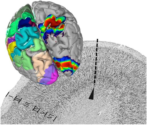
Under the microscope, it can be seen that the human brain is not uniformly structured, but divided into clearly distinguishable areas. They differ in the distribution and density of nerve cells and in function. With the Julich-Brain, researchers led by Katrin Amunts now present the most comprehensive digital map of the cellular architecture and make it available worldwide via the EBRAINS research infrastructure.
Forschungszentrum Jülich / Katrin Amunts
The architecture of the nerve cells changes at the border between two areas (dotted line). This is the basis for mapping. The areas of brains
|
"On the one hand, the digital brain atlas will help to interpret the results of neuroimaging studies, for example of patients, more accurately", says Katrin Amunts, Director at the German Research Center Juelich and Professor at the University of Düsseldorf. "On the other hand, it is becoming the basis for a kind of 'Google Earth' of the brain - because the cellular level is the best interface for linking data about very different facets of the brain.
A Google Earth of the Brain
In this way, the researchers are making a significant contribution to the Human Brain Project (HBP), for which the European Commission just approved 150 million Euro until 2023. "Together with many partners in this project, we are building EBRAINS as a novel high-tech research infrastructure for the neurosciences," says Amunts, who is also the Scientific Research Director of the project.

Prof. Katrin Amunts is Director of the Institute of Neuroscience and Medicine at Forschungszentrum Jülich and Professor of Brain Research at University of Düsseldorf in Germany
More than a quarter century of research has gone into the 3D atlas. Dozens of experts have used image analysis and mathematical algorithms to evaluate the tissue sections over the years and determine the boundaries between brain areas, which together make up a length of almost 2000 metres.
Regions vary in their difference
Mapping showed that areas vary between different brains, for example in terms of size and location. The Julich-Brain therefore displays the position and shape of individual regions as "probability maps". The researchers found particularly large differences in the Broca region, which is involved in language. In contrast, the primary visual area appeared much more uniform.
As part of EBRAINS, the Jülich Brain Atlas is the starting point for bringing structure and function together. The atlas is already helping to link data on gene expression, connectivity and functional activity, for example, to better understand brain functions and the mechanisms of diseases. "EBRAINS also enables us to use the maps for simulations or to apply artificial intelligence to explore the division of labor between brain areas. The huge amounts of data generated from this are processed using the EBRAINS computing platform." The computational power comes from the new European supercomputing network FENIX, which is formed by five leading centres for High Performance Computing, including the Jülich Supercomputing Centre (JSC).
Digital brain science
"It is exciting to see how far the combination of brain research and digital technologies has progressed," says Amunts. "Many of these developments converge in the Julich-Brain-Atlas and on EBRAINS. They help us – and more and more researchers worldwide – to better understand the complex organization of the brain and to jointly uncover how things are connected.”
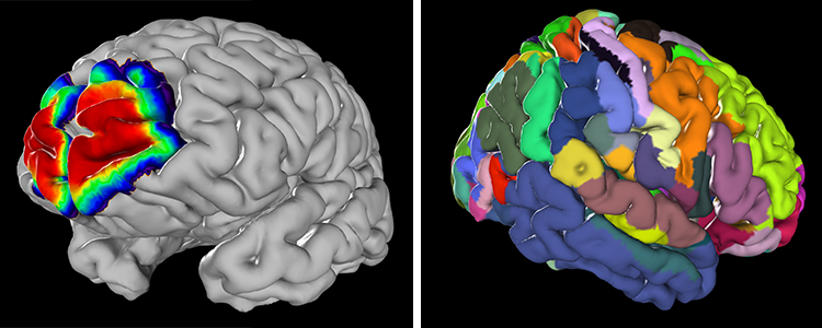
Probability map of the frontal pole, and overall map view. Forschungszentrum Jülich / Katrin Amunts
Original publication:
Katrin Amunts, Hartmut Mohlberg, Sebastian Bludau, Karl Zilles: Julich-Brain - a 3D probabilistic atlas of human brain's cytoarchitecture. Science (First Release)
DOI: 10.1126/science.abb4588
BigBrain
The high-resolution brain model BigBrain, published in Science in 2013, is included in the Julich Brain Atlas and also available via EBRAINS. It is currently being investigated using new AI-based methods by the German-Canadian HIBALL lab, a partnering project of the HBP.
Contact:
Prof. Katrin Amunts
k.amunts@fz-juelich.de
Footage for download:
Web-based access to the atlas functionalities at EBRAINS (mp4)
Laboratory footage with co-author Sebastian Bludau (mp4, royalty-free. Source: European Commission Stockshots)
See also:
Linking gene expression to brain microstructure