
Young Researchers Event 2019
EBRAINS - A platform for collaboration in digital neuroscience
9 July 2019 | Belgrade, Serbia
FAQs & ALL YOU NEED TO KNOW
Belgrade by plane
Belgrade’s main airport is Nikola Tesla Airport, located in Surčin, 18 km away from Belgrade’s city centre. There are direct flights from most major European cities as well as connections also form Asia, Africa and North America.
Belgrade by train
The centrally located Belgrade Centre Railway Station (Beograd Centar Prokop) is connected to all main cities of Europe by daily and overnight trains. There are commercial discounts available as Euro Domino, Railplus, Balkan Flexipass, Easy Travel Card, Euro 26, City Star and the unified Balkan Tariff. Find further info here: Serbian Railways
Belgrade by car
The main international roads going through Belgrade are E-70 and E-75. It is necessary to pay on several tolls on your way to Belgrade. As the local roads may lack of sufficient road signs, you should use a navigating system.
Driving speed:
- In town: 50 km/hour.
- Outside built-up areas: 80-100 km/hour
- Motorways: 120-130 km/hour.
Airport transfer
It is safer to book a transfer in advance, but if this is not possible, you have following possibilities:
- Public transport
The cheapest possible transfer solution is certainly a regular bus, specifically line 72 which takes up to 60 minutes and costs around 1 €. There are also minibuses available, which take you 30 to 40 minutes to Belgrade centre and cost around 2 €.
- Taxi
Taxis are easily available at Belgrade airport. There is also a city service TAXI INFO desk where you can take a taxi receipt with the name of your destination and appropriate pricing for the taxi service.
The average taxi’s price to the city center is around 15 € and depending on traffic takes you 20-30 minutes.
Public transport
Belgrade’s public transport consists of a network of bus, trolleybus and tram routes. Tickets can be bought in the transport vehicle from the driver or conductor as well as at kiosks that are marked with a ticket sales sticker. The bus lines run every 3 to 30 minutes at daytime, whereas the night lines run every 30-60 minutes.
The closest tram/bus stop is called “Karađorđev Park” (Карађорђев парк).
In order to get to the venue from the main train station, you need to do the following:
-
Take bus 78 or Tram 9 (direction Banitsa) and get off at the named stop (should be the 4th stop).
-
Walk against the direction you came from and turn left on the first corner. Then you need to turn left again and walk along the street “Dr Subotića starijeg (др Суботића старијег)” until you see the university’s building on your right.
-
The walk from the bus station to the venue is about 5 minutes. You can also walk from the Train station, as this would also take you only 12 minutes.
By taxi
Telephone: +38111 19804 further information
Mainly there are two different Taxi rates:
-
From 6 am until 10 pm it starts around €1,40 and the kilometre charge is 50cent.
-
From 10pm until 10am the rate also starts around €1,40, but the charge per kilometre is around 70c.
Taxis can also be stopped on the street but be careful to not catch an unlicensed taxi. In order to prohibit this, you should make sure that the taxi has a company board or unique number on the roof, a taximeter and a registration plate with the letters TX at its end.
The closest accommodation facilities to the venue of the event are the ones in Vracar and Savski Venac districts. Here are some suitable hotels:
Belgrade has a moderate continental climate, with hot humid summers and cold winters. In the time from April-October you find the sunny months in Belgrade. The daily temperature in July will be between 17 and 28° which results in a daily mean of 23°.
- Language
The main language in Belgrade is Serbian. But English is widely spoken as well. Especially in restaurants, bars, shops and other public places in the city center, you can perfectly communicate with English. Serbian belongs to the Slavic languages and besides the latin script, Serbian is also written in Cyrilic.
- Currency, credit cards, banking
The official currency is Serbian Dinar (RSD, РСД). The exchange rate is around: 1 € = 117,60 RSD. For your exchange of money, you should turn to specialized banks or one of the many exchange offices. Exchange machines accept Euro, US dollars and British pounds. Dinar is used in denominations of 10, 20, 50, 100, 200, 500, 1000 and 5000. The latest exchange rate from Euro to Dinar can be checked here. Of course you can purchase your money also directly from an ATM, which you usually find in major malls and public places. Most international credit cards (Eurocard/Mastercard, Visa, American Express) are also widely accepted.
- Time zone
Belgrade is in the Central European Time Zone (CET). In the winter months clocks are set at GMT + 1 hour and in the summer (March to end of October) GMT + 2hours.
- Electric current
Electric sockets in Serbia have a voltage of 220 Volts and a frequency of 59Hz. Adapters may be needed.
Please note that the information provided on this site has been obtained from several different sources and therefore the organisers cannot accept any responsibility for errors therein.
The human brain is a multi-level and highly complex system that produces, processes and transmits information in an unique manner. Researchers and scientists from all over the world strive to decode the mechanisms underlying this unique system. The European Human Brain Project plays a pioneering role in neuroscience research, uniting experts from various fields in the aim to build a collaborative platform for computational neuroscience.
This event was open to the entire scientific community but especially targeted early-career researchers. The programme provided an overview of interdisciplinary efforts towards collaboration for the advance of digital neuroscience and offered ample opportunities for participants to exchange information and knowledge with peers as well as renowned experts.
Participation in the event was free.
SCIENTIFIC PROGRAMME
The scientific programme is also available as PDF download:
![]() Programme - Young Researchers Event (1.7 MB)
Programme - Young Researchers Event (1.7 MB)
PROGRAMME
Welcome and Introduction to the European Human Brain Project | 20 min
Katrin Amunts (Forschungszentrum Jülich)
EBRAINS – towards a European research infrastructure | 30 min
Jan Bjaalie (University of Oslo)
The visual cortex: When structure meets function | 30 min
Aleksandar Maliković (University of Belgrade)
Introduction to the HBP Collaboratory | 20 min
Jonathan Villemaire-Krajden (EPFL)
Introduction to the HBP Knowledge Graph | 20 min
David Kunzmann (EPFL)
Micronanatomy of the brain: A critical step to model neuronal circuits | 15 min
Marta Turégano (Universidad Politécnica de Madrid)
Ethics in neuroscience research | 15 min
Simisola Akintoye (De Montfort University)
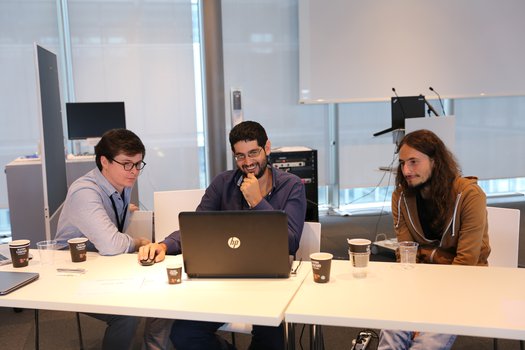
Hands-on sessions | 2 x 90 min
- Hands-on session I: Open Science: How to
Camilla Hagen Blixhavn & Jan Bjaalie (University of Oslo)
- Hands-on session II: Simulation in neuroscience
Alexander Peyser (Forschungszentrum Jülich)
- Hands-on session III: The HBP Human Brain Atlas: Integration and visualization of heterogeneous neuroscience data
Lyuba Zehl & Kai Kiwitz (Forschungszentrum Jülich, Heinrich Heine University Düsseldorf)
Hands-on career development: Insights into supportive measures of the HBP | 15 min
Facilitator: Karin Grasenick (convelop cooperative knowledge design gmbh)
Round-table discussion: Feedback session on HBP Infrastructure | 30 min
Young researchers: Isidora Banjac, Radmila Perić, Joko Poleksić (University of Belgrade)
Senior researchers: Katrin Amunts, Jan Bjaalie, Aleksandar Maliković
Cognition and motion: Space to be fulfilled
(Guest lecture - open to the public) | 30 min
Vladimir Kostić (University of Belgrade)
CONFIRMED SPEAKERS
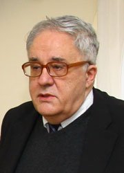
 Jan Bjaalie, M.D., Ph.D., is professor at the Institute of Basic Medical Sciences, University of Oslo, and Infrastructure Operations Director and leader of the Neuroinformatics Platform of the EU Human Brain Project. He was founding Executive Director of the International Neuroinformatics Coordinating Facility (INCF) and is currently head of the INCF Norwegian Node and member of the INCF Council for Training, Science, and Infrastructure. His research group has studied wiring patterns of the brain and developed data systems for organizing and managing heterogeneous neuroscience research data by use of a new generation of digital brain atlases. The group develops software and workflows for analysis of data integrated in the atlases. Jan Bjaalie is Chief editor of Frontiers in Neuroinformatics and Section editor of Brain Structure and Function.
Lecture title: EBRAINS - towards a European research infrastructure
No abstract provided.

Simisola Akintoye is the Interim Data Protection Officer for the European Union Future and Emerging Technologies Human Brain Project. She is an international privacy practitioner, data protection consultant and law lecturer at De Montfort University Institute of Evidence-based Law Reform where she is the convenor of Privacy, Ethics and Responsibility.
Her work involves continuous, up-to-date collaborative research in privacy and data protection. As a legal expert, she regularly sits on panels involved in critical dialogues on balancing the competing interests of privacy and innovation at national and international levels. She holds a Phd in Law – Corporate Governance and Regulation in Emerging Economies (Sheffield), LLM International Commercial Law (Dundee), LLM Business Law and Taxation (France), Certified Data Protection Practitioner (PC.dp) on European Union General Data protection Regulation (EU GDPR) from Privacy and Data Protection (PdP) London.
Lecture title: Ethics in neuroscience research
No abstract provided.

Katrin Amunts did a postdoctoral fellowship at the C. & O. Vogt Institute of Brain Research at Düsseldorf University, Germany. In 1999, she set up a new research unit for Brain Mapping at the Research Center Juelich, Germany. In 2004, she became professor for Structural-Functional Brain Mapping, and in 2008 a full professor at the Department of Psychiatry, Psychotherapy and Psychosomatics at the RWTH Aachen University as well as director of the Institute of Neuroscience and Medicine (INM-1) at Research Center Juelich. Since 2013, she is a full professor for Brain Research, director of the C. and O. Vogt Institute of Brain Research, Heinrich-Heine University Düsseldorf and director of the Institute of Neuroscience and Medicine (INM-1), Research Center Juelich.
Katrin Amunts is a member of the editorial board of Brain Structure and Function. She is a member of the German Ethics Council since 2012, and has been elected vice chair in 2016. Katrin Amunts is the programme speaker of the programme Decoding the Human Brain of the Helmholtz Association, Germany. She is leading Subproject 2 Human Brain Organisation of the European Flagship Project The Human Brain Project (HBP). In 2016, she has been elected Scientific Research Director and Chair of the Science and Infrastructure Board (SIB) of the HBP. Since 2017 Katrin Amunts is co-speaker of the graduate school Max-Planck School of Cognition and since 2018 she is a member of the International Advisory Council Healthy Brains for Healthy Lives, Canada.
In order to better understand the organisational principles of the human brain, she and her team aim to develop a multi-level and multi-scale brain atlas, and use methods of high-performance computing to generate ultra-high resolution human brain models.
Lecture title: Welcome and introduction to the European Human Brain Project
The human brain has a multi-level organisation and high complexity. New approaches are necessary to decode the brain with its 86 billion nerve cells, which form complex networks. Ultra-high resolution models and simulation pose massive challenges in terms of data processing, visualisation and analysis. For example, high-resolution data obtained in post-mortem brains supplement information about structural and functional connectivity as obtained by Magnetic Resonance Imaging, which can be acquired from the living human brain, with a spatial resolution in the millimetre range. 3D Polarized Light Imaging elucidates the connectional architecture of the brain at the level of axons at the micrometre range, while keeping the topography of the whole organ; it results in data sets of several petabytes per brain, which should be actively accessible while minimizing their transport. By bringing together different aspects of connectivity in one and the same atlas, it is feasible to combine the advantages of the different approaches, and to address the different spatial (and temporal) scales.
The Human Brain Project is developing a multimodal human brain atlas, which integrates the information of brain organisation from multiple levels including cellular architecture, connectivity, molecular and genetic maps, and results from neuroimaging studies of the brain from healthy subjects and patients. Empirical research is supplemented by modelling and simulation, resulting in new data that is stimulating new experiments.
The Human Brain Project [1, 2] creates a cutting-edge European infrastructure to enable cloud-based collaboration among researchers coming from different disciplines around the world, to create development platforms with databases, workflow systems, petabyte storage, and supercomputers opening new perspective to decode the human brain.
References
[1] K. Amunts, A. Knoll, T. Lippert, C.M. Pennartz, P. Ryvlin, A. Destexhe, V.K. Jirsa, E. D’Angelo, J.G. Bjaalie, The Human Brain Project – synergy between neuroscience, computing, informatics and brain inspired technologies, PLoS Biol, in press (2019).
[2] A. Salles, J.G. Bjaalie, K. Evers, M. Farisco, B.T. Fothergill, M. Guerrero, H. Maslen, J. Muller, T. Prescott, B.C. Stahl, H. Walter, K. Zilles, K. Amunts, The Human Brain Project: Responsible brain research for the benefit of society, Neuron, 101 (2019) 380-384.
 Karin Grasenick graduated in Computer Science and Biomedical Engineering. Her thesis on innovative non-invasive techniques to measure stroke volume led to a growing interest in inter- and transdisciplinary research, diversity in research content and equal opportunities in science. She lectures, coaches and supports teams, universities and international projects in diversity and change management. In HBP, she actively supports the recognition of diversity as a success factor for research and innovation. Together with her team she provides tools and techniques for researchers accordingly.
Lecture title: Hands-on career development: Insights into supportive measures of the HBP
Keynote Speakers will give insights on success factors and challenges for scientific careers and answer questions of participants. General rules and differences will be explored in the context of equal opportunities in the global scientific community. Specific focus will be set on HBP best practices and measures to support career development regardless of gender and further diversity traits.
 Aleksandar Maliković
is a Full Professor of Anatomy at the Faculty of Medicine at the University of Belgrade and his research focus is on the brain mapping and the architectonics of human visual cortex. He is a doctor of medicine (M.D.), Master of Medical Science (M.Sc.) and Doctor of Science (D.Sc. or Ph.D.). Aleksandar was awarded fellowship of the European Federation for Experimental Morphology (1996) and Heinrich Heine University (1997), C. and O. Vogt Institute of Brain Research, Dϋsseldorf, Germany. He spent 2 years as Visiting Fellow at C. and O. Vogt Institute of Brain Research, Heinrich Heine University, Dϋsseldorf, Germany under the supervision of Prof. Karl Zilles who had a strong influence on him and who created many opportunities for him. Also, he spent 2 years working as Visiting Scientist at the Research Centre Jϋlich, Institute of Neuroscience and Medicine (INM-1) - Structural and Functional Organization of the Brain where he collaborated with Prof. Katrin Amunts. Aleksandar is leading a project Structure and function of the Visual System (Ministry of Education, Science and Technological Development, Serbia) and he was a collaborator in the following projects: Human Brain Model (Helmholtz Gemeinschaft, Germany), Grant 01GO0204 (Brain Imaging Center West, Bundesministerium für Bildung und Forschung, Germany), Human Brain Project/Neuroinformatics (National Institute of Biomedical Imaging and Bioengineering, National Institute of Neurological Disorders and Stroke and National Institute of Mental Health, USA) and Morphological Investigations of the Brain (Ministry of Education, Science and Technological Development, Serbia). He is a member of Serbian Anatomical Society, Serbian Neuroscience Society, Federation of European Neuroscience Societies, European Federation for Experimental Morphology (board member) and Anatomische Gesellschaft. Aleksandar is the author or co-author of approximately 50 peer-reviewed articles, 3 monographs, 9 textbooks and 11 anatomical atlases.
Lecture title: The visual cortex: When structure meets function
Considerable technological progress in noninvasive functional investigations of the human visual cortex enabled the discovery of new visual areas and their further precise functional mapping. Since the beginning of the 1990s, the use of functional magnetic resonance imaging (fMRI) and positron emission tomography (PET) enabled the functional mapping of 13 occipital visual areas: V1, V2, V3 (V3d/V3v), V3A, V4, VО1, V5, hMST, V6, V7, LO1, LO2. This significant scientific progress in the functional mapping of the human visual cortex was not followed by the corresponding architectonic investigations so it was not possible to establish anatomical correlates of these new functionally defined visual areas. During the past two decades, our enthusiastic and dedicated group performed cytoarchitectonic mapping of 11 occipital areas (hOc1, hOc2, hOc3v, hOc4v, hOc3d, hOc4d, hOc4la, hOc4lp, hOc5, FG1 and FG2) which are visual in their function. Continuous probabilistic maps of these cytoarchitectonic areas have been generated to characterise their location, extent, size and variability. These maps are easily available to the whole scientific community (JuBrain, Human Brain Project) and their verification is ongoing. Convergent evidence from a standpoint of location, topography, size, functional properties and connectivity suggests that these cytoarchitectonic areas are the anatomical correlates of the functionally defined visual areas.
No bio and pic provided.
Lecture title: Hands-on session II: Simulation in neuroscience
No abstract provided.
 Vladimir S. Kostić, Professor of Neurology at the Medical School of Belgrade, has focused his scientific interest in degenerative disorders of the central nervous system, mainly etiopathogenesis and treatment. In the course of his training and subsequent academic carrier he worked apprx. 3 years in the Neurological Institute of the Columbia Univesity (New York; USA). He is the author of 7 textbooks and editor of 12 monographs. He is also the author or co-author of >300 peer-reviewed papers. He is heading several scientific projects mainly devoted to the genetic background of neurodegenerative disorders. He is the member of the Serbian Academy of Science and Arts and Honorary President of the Serbian Neurological Society.
Lecture title: Cognition and motion: Space to be fulfilled
In several studies, including one recruiting 28,808 community-dwelling adults, it has been shown an association between poor cognitive function and reduced mobility (gait, balance and chair stands). Such interdependence between mobility and cognition has been suggested to reflect age-dependent changes in shared neural mechanisms and an increased demand for cognitive monitoring in motor control with age. Indeed, safe and independent negotiation of complex environments requires integration of external sensory information with neural networks involving cortical, subcortical, brainstem, and spinal cord structures (some of these share common substrates with cognition). A substantial corpus of evidence suggests that the cognitive involvement in postural control and gait increases with aging. A large portion of such studies were based on dual-task experimental designs, which typically use the simultaneous performance of a motor task (e.g., static or dynamic balancing, walking) and a continuous cognitive task (e.g., mental arithmetic, tone detection). Resolution of similar interferences requires executive functions, which schedule processing order of the tasks, interrupt or reinstale task processing, and switch between the processing streams of the tasks. Even in cognitively normal, older adults, there is a robust association of gait speed and gait variability with executive function, attention, and (to a lesser extent) memory and visuospatial function. These associations are more emphatic under dual-task conditions. Moreover, gait impairment precedes and predicts cognitive decline and may be a more sensitive marker of cognitive decline than cognitive tests.
We have examined this association in patients with Parkinsonʼs disease (PD) using gait and freezing of gait (FoG) as models. In PD patients during the mental dual-task we observed furher deterioration in gait parameters which reflect gait automaticity (eg. an increase in stride variability and gait asymmetry, in parallel with decrease in gait rhythmicity). In PD patients with FoG we conducted multimodal MRI investigations and found (a) functional decoupling between coordinated motor and cognitive neural networks (“interference model” of FoG) and (b) altered functional connectivity not only in the locomotor but also in “cognitive” circuits such as the attention-related frontoparietal and visual occipito-temporal networks („network-wide dysfunction“).

Camilla Hagen Blixhavn is a researcher at the University of Oslo and team coordinator for atlas integration of rodent brain research data in the Human Brain Project. She couples her experience from organisational and creative leadership positions together with her studies in psychology, biology, didactics and neuroscience, and continues to strategise towards the needs of researchers in science and health. Camilla is MSc in neuroscience and is preparing for an interdisciplinary PhD in neuroscience data management.
Lecture title: Hands-on session I: Open Science: How to
Open Science is a continuation of practices which began centuries ago with the advent of academic journal publications. The most recent developments in Open Science is Open Data, recently made possible by new technological capabilities to store large amounts of data, and to access and work on the data online. Leading journals are already now requiring posting of research data in public repositories, to complement the traditional journal article containing the interpretations of the data. Several general data repositories for sharing of research data are available – however, many lack the necessary stewardship and standards for making the research data easy to Find, Access, Interpret, and Re-use, in accordance with the FAIR principles (Wilkinson et al., Scientific Data 3:160018, 2016). To address these challenges, the Human Brain Project is developing a new research infrastructure, the EBRAINS FAIR data service, providing tools, workflows and data curation services tailored for integration and sharing of heterogeneous, multimodal neuroscience data and computational models. Three-dimensional open-access brain reference atlases provide the anatomical context for all data shared via the EBRAINS infrastructure, easing comparison and interpretation of findings.
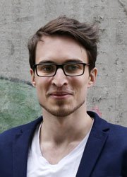
 Formerly involved in the music industry, David Kunzmann decided to change paths in 2013 and study Computer Science. After gaining experience in diverse startups, he joined the HBP Knowledge Graph team at the end of 2017 as a full stack developer.
Lecture title: Introduction to the HBP Knowledge Graph
Open science is all about making the scientific data that is produced findable, accessible, interoperable and re-usable by others. Discover how the Human Brain Project annotates its heterogeneous research data with metadata in the HBP Knowledge Graph and how this allows you to find and re-use existing high-quality research data and/or share your own.
Jonathan Villemaire-Krajden is a developer who has been with the Human Brain Project since 2018. He works on the Collaboratory tools and infrastructure with a few colleagues in Geneva, Switzerland. He grew up and studied in Canada as a computer scientist and physicist and worked as a developer and devops engineer since 2010. He is interested in science, the outdoors and software design.
Lecture title: Introduction to the HBP Collaboratory
The Human Brain Project (HBP) Collaboratory is a central directory for tools developed by the HBP Platforms. At the core of the Collaboratory are Collabs. These are spaces designed for collaborative work where users can contribute content, integrate applications and run Jupyter notebooks. Collabs have at least a team, a landing page, and a navigation menu. As a user, you can organise content, which can be text or documents, manage the team members you want to work with and add interactive tools. The Collaboratory is a tool to help with the dissemination of science, collaboration, and project management. In this presentation, I will introduce the project and the vision behind it, show you where to find information, give a demo of the HBP Collaboratory's features and present how to build a shared workspace for a simple project.

Lyuba Zehl studied Biology (BSc) and Neuroscience (MSc) at the University of Cologne under the supervision of Prof. Ansgar Büschges and Prof. Wolfgang Walkowiak, respectively. During this time of study, she worked on kinematic of insect legs during locomotion (BSc thesis), optimisation of peripheral nerve implantation of multi-electrode arrays in small mammals (internship at Blackrock Microsystems and the Center of Neural Interfaces of the University of Utah, USA), and the anatomical cartography of auditory nuclei in the brain stem of toothed whales using cluster analysis (MSc thesis). For her doctoral studies, she switched to RWTH Aachen University and joint the Statistical Neuroscience Group of Prof. Sonja Grün at the Institute for Neuroscience and Medicine (INM-6) of the Jülich Research Centre. In her thesis, she worked on analysing multi-electrode array recordings of monkey motor cortex and, in particular, on data and metadata management of complex neuroscience experiments. After receiving her doctorate degree (Dr. rer. nat.) in 2017, she started working as a junior scientist half-time under Timo Dickscheid in the Big Data Analytics Group at the INM-1 of the Jülich Research Centre and Molecular and half-time under Prof. Björn Kampa in the Systemic Neurophysiology Group at the Institute of Biology II at RWTH Aachen University. Since May 2019, she has worked full time in the Big Data Analytics Group at the INM-1 for the curation team of the Neuroinformatics Platform of the Human Brain Project (HBP). Having a high interdisciplinary orientation towards computational neuroscience and software development for data and metadata management and a broad experience in various neuroscience laboratories, she now focuses on developing and implementing concepts, standards and tools for neuroscience data and metadata management. With her current work, she strongly supports the integration of heterogeneous neuroscience data into the unified data sharing platform and the interactive atlas viewers of the HBP.
Lecture title: Hands-on session III: The HBP Human Brain Atlas: Integration and visualisation of heterogeneous neuroscience data
As a part of the Neuroinformatics Platform, the Human Brain Project (HBP) is developing a comprehensive multi-level Human Brain Atlas. This novel atlas consists of web-based services that allow users to discover, explore and access a unique selection of anatomical reference spaces and high-quality structural parcellations, down to a microscopic resolution. A diverse range of well curated human brain data at different scales and modalities is linked to this atlas, and can be discovered and downloaded by browsing brain regions. For integrating new data to the atlas, the platform provides support and tools for metadata curation, and spatial anchoring of non-image, 2D and 3D image data to the reference atlases. A rich interactive viewer allows users to display common types of data in anatomical context together with the reference atlas delineations (e.g., JuBrain cytoarchitectonic atlas, BigBrain microscopic model). During the hands-on session, participants learn how the BigBrain reference model can be interactively explored in 3D using the interactive atlas viewer, and how non-image data can be semantically linked to the JuBrain reference atlas, using cortical receptor densities as an example. We will also demonstrate spatial anchoring of 2D and 3D image data to the HBP’s reference atlases.
Kai Kiwitz graduated in Psychology and Cognitive Neuroscience at the Universities of Düsseldorf and Bochum in 2016. Since then, he performs his PhD work at the Cécile and Oskar Vogt Institute of Brain Research at the University of Düsseldorf under the supervision of Prof. Katrin Amunts in close collaboration with the Big Data Analytics Group led by Dr. Timo Dickscheid at the INM-1 of the Jülich Research Centre. In his research, Kai evaluates the use of machine learning solutions aimed at aiding neuroscientists in high-resolution cytoarchitectonic mapping approaches in the BigBrain. He is especially interested in analysing representations of cytoarchitectonic features within Convolutional Neural Networks to validate their use in the generation of a high-resolution anatomical reference framework of the human visual system.
Lecture title: Hands-on session III: The HBP Human Brain Atlas: Integration and visualisation of heterogeneous neuroscience data
As a part of the Neuroinformatics Platform, the Human Brain Project (HBP) is developing a comprehensive multi-level Human Brain Atlas. This novel atlas consists of web-based services that allow users to discover, explore and access a unique selection of anatomical reference spaces and high-quality structural parcellations, down to a microscopic resolution. A diverse range of well-curated human brain data at different scales and modalities is linked to this atlas, and can be discovered and downloaded by browsing brain regions. For integrating new data to the atlas, the platform provides support and tools for metadata curation, and spatial anchoring of non-image, 2D and 3D image data to the reference atlases. A rich interactive viewer allows users to display common types of data in anatomical context together with the reference atlas delineations (e.g., JuBrain cytoarchitectonic atlas, BigBrain microscopic model). During the hands-on session, participants learn how the BigBrain reference model can be interactively explored in 3D using the interactive atlas viewer, and how non-image data can be semantically linked to the JuBrain reference atlas, using cortical receptor densities as an example. We will also demonstrate spatial anchoring of 2D and 3D image data to the HBP’s reference atlases.

Marta Turégano is a PhD student at the Cajal Laboratory of Cortical Circuits directed by Prof. Javier DeFelipe in Madrid (Spain). She holds a degree in Biology and a Master’s degree in Neuroscience by the Universidad Autónoma de Madrid. During those stages she learnt 3D electron microscopy techniques applied to the exploration of the ultrastructure of the brain. Currently, her research is focused on the description of the mouse somatosensory cortex, the synapses, axons, dendrites and organelles placed in these neuronal compartments. Her work is part of the subproject 1 “Mouse Brain Organisation” of the Human Brain Project. In addition, she is the SP1 Student Ambassador, in which function she promotes the events of HBP aimed at the student community.
Lecture title: Microanatomy of the brain: A critical step to model neuronal circuits
Most cortical synapses are established in the neuropil, which is composed of dendrites, axons and glial processes. There are two basic types of synapses: excitatory (glutamatergic) and inhibitory (GABAergic). It is important to describe the morphology of both kind of synapses, as well as their density and location, because these parameters have a functional correlate. In addition, we have studied the organelles placed on these neuronal compartment as mitochondria, spine apparatus and multivesicular bodies (MVBs) and their possible relation with synapses. For that purpose, we used three-dimensional electron microscopy with combined focused ion beam milling and scanning electron microscopy (FIB/SEM), a method that allows us to obtain long series of consecutive sections in an automated way. These stacks of serial sections can later be examined and segmented in 3D with dedicated software called Espina. We have created a new tool of Espina to trace the fibers across the stacks of EM images, acting as the skeletons of different compartments in the sample. Besides qualitative information, we extract morphological information about the length, density and connectivity of dendrites, axons and the synapses among them.
This work provides accurate quantitative data that helps understand the mouse cortical circuitry and will be very useful to refine and validate current cortical models.
SCIENTIFIC CHAIRS
Katrin Amunts | HBP Scientific Research Director
Jan Bjaalie | HBP Infrastructure Operations Director
Nebojša Lalić | Dean University of Belgrade, Faculty of Medicine
Tatjana Pekmezović | Vice Dean University of Belgrade, Faculty of Medicine
Aleksandar Maliković | University of Belgrade, Faculty of Medicine
PROGRAMME COMMITTEE
Katrin Amunts | Forschungszentrum Jülich, Heinrich Heine University Düsseldorf, Germany
Jan Bjaalie | University of Oslo, Norway
Aleksandar Maliković | UBFM, Serbia
Sandra Diaz | Forschungszentrum Jülich, Germany
Martina Schmalholz | Heidelberg University, Germany
Gabriel Urbain | Ghent University, Belgium
ORGANISERS
HBP Education Programme Office
education@humanbrainproject.eu
![]()
ABOUT THE VENUE
UNIVERSITY OF BELGRADE
Faculty of Medicine
Dr Subotića 8
11 000 Belgrade
Serbia
The Young Researchers Event 2019 will take place at the University of Belgrade, Faculty of Medicine (UBFM). The venue offers a big ceremonial room where all the main agenda points of the event will take place. Additionally, there are two smaller rooms that will be used for hands-on sessions.
The University of Belgrade is the leading institution of higher education in the region. Its Faculty of Medicine is only 12 minutes of walking from the main train station of Belgrade (Beograd Centar-Prokop).