
New Cytoarchitectonic maps in BigBrain
03 June 2019
Maps of cytoarchitectonic areas and cortical layers released for BigBrain
The BigBrain is a microscopic resolution 3D model of the human brain, reconstructed from digital scans of thousands of histological sections (Amunts, et al., Science, 2013) through a collaboration between the teams of Katrin Amunts at Forschungszentrum Jülich and Alan Evans at McGill University, Montreal.
With an isotropic resolution of 20 micron, it is the most detailed human brain model available to date, and one of three reference templates of the HBP atlases. HBP has now released a first set of 3D maps of cytoarchitectonic areas for the BigBrain, which are based on expert delineations employing image analysis and multivariate statistics in individual histological, coronal sections of the BigBrain.
To extend the expert delineations into gapless 3D maps, different strategies have been employed: For two areas of the visual system - hOc1 (BA17/V1 in the CalcS) and hOc2 (BA18/V2) - a Deep Learning algorithm has been used to complement the expert annotations by detailed automatic delineations in every single histological section (total number of mapped sections for hOc1: 2430 and hOc2: 2454), leading to highly detailed 3D maps.
For several other areas, preliminary 3D maps are provided based on direct interpolation of the expert delineations, which were made in every 15th to 60th section over the whole extent of the areas (see Fig. 1). These areas include TE 1.0 (HESCHL), TE 1.1 (HESCHL), TE 1.2 (HESCHL), TE 3 (STG), STS1 (STS), STS2 (STS), 6d1 (PreCG), 6d2 (PreCG), 6ma (preSMA, mesial SFG), 6mp (SMA, mesial SFG), 6d3 (SFS), ifj1 (IFS/PreCS), ifj2 (IFS/PreCS), ifs1 (IFS), ifs2 (IFS), ifs3 (IFS), ifs4 (IFS), Entorhinal Cortex, hIP4 (IPS), hIP5 (IPS), hIP6 (IPS), hIP7 (IPS), hPO1 (POS), and hOc6 (POS).
The preliminary maps will be updated incrementally by highly detailed versions in the future, and more and more new areas will be added.
Complementing these maps of cytoarchitectonic areas, HBP also provides access to a new cortical layer segmentation in the BigBrain (Wagstyl et al. 2019), which provides detailed whole-brain maps and surface meshes of 6 cortical layers (see Fig. 2).
All these maps can be explored interactively in HBP’s 3D atlas viewer at http://bigbrain.humanbrainproject.eu, and found in HBP's „Knowledge Graph Search“ (https://www.humanbrainproject.eu/en/explore-the-brain/search/) for detailed information and download.
FIGURES:
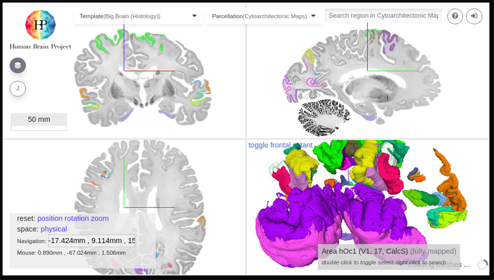 Fig. 1: Preliminary 3D maps based on direct interpolation of expert delineations.
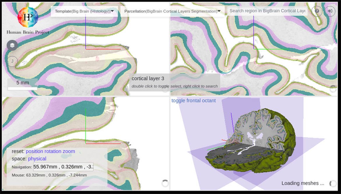 Fig. 2: Cortical layer segmentation which provides detailed whole-brain maps and surface meshes of 6 cortical layers.
ENDS