
- Press Release
Brain modelling used to identify necessary circuits of consciousness
16 June 2023
Researchers of the Human Brain Project have used a model-based approach to identify the brain circuits implicated in consciousness. The results of the study, a collaboration between Pompeu Fabra University in Barcelona and University of Liège, have been published in the journal Human Brain Mapping. The team studied the propagation of signals in models of the brain of patients with disorders of consciousness (DoC), identifying two relevant circuits in the posterior cortical region and the thalamo-frontotemporal region. The results bring more understanding of the inner workings of brain networks and could improve diagnosis and even provide treatment targets for people suffering from DoC.
Currently, patients with DoC are classified in several categories (coma, unresponsive wakefulness syndrome and minimally conscious state) which describe their overall consciousness and awareness. “The diagnosis is mainly response-based: the doctor sits down with the patient and assesses their response to stimuli” explains Jitka Annen from the University of Liège. “However, this may not correspond to their underlying brain activity - patients with high activity may still be unable to react. It’s a heterogenous group. We wanted to go a step beyond assessment and classification based on neuroimaging and instead look at the flow of information in their brains, to find common patterns associated with consciousness”.
The researchers focused on two DoC groups - unresponsive wakefulness syndrome (previously known as vegetative state) and minimally conscious state. After collecting fMRI data from each patient during resting state (i.e., patients were awake but no particular task was provided), they looked at spontaneous and model-based perturbation of brain activity captured by the blood flow, such as signals and peaks. “Based on spontaneous peaks of activity, we evaluated the personalised connectivity of each patient’s brain, which can tell us how likely a signal is to travel from one point to the other” says Gorka Zamora-López from Pompeu Fabra. “After we constructed a patient-specific computational model of their propagation patterns, we then trigger a signal in the model and see how the brain reacts. In particular we look for which areas are more likely to respond to a signal; which areas are more likely to propagate it. Basically, we look at whether an area acts as influencer or influenced”.
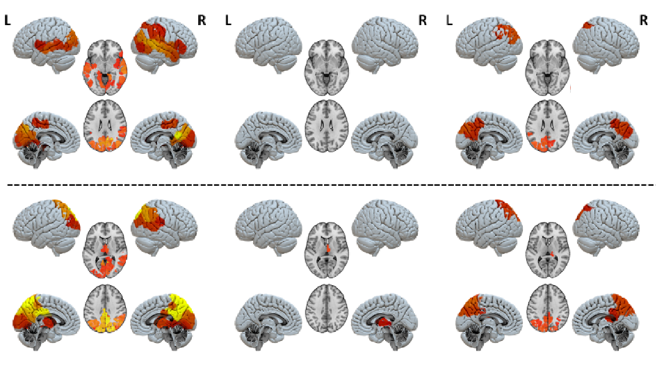
A marked distinction arises between the unresponsive wakefulness syndrome group and minimally conscious state group, with the former not displaying activity in identifiable circuits. “The key difference is that in patients with unresponsive wakefulness syndrome no region of the brain seems embedded in a functional network, they all display equally low activation. Meanwhile, distinct regions and circuits pop out in the brain models of people in minimally conscious state: the thalamo-frontotemporal region when broadcasting signals, and the posterior cortical region when receiving them” adds Rajanikant Panda of the University of Liège.
These findings shed new light on disorders of consciousness, and could lead to a more defined understanding of the mechanisms based on brain activity rather than behavioral responses. “I believe these results can potentially guide the clinicians to better understand what is going wrong in the information exchange and thus look for ways to reactivate those circuits” concludes Zamora-López.
Text by Roberto Inchingolo
Reference: Rajanikant Panda, Ane López-González, Matthieu Gilson, Olivia Gosseries, Aurore Thibaut, Gianluca Frasso, Benedetta Cecconi, Anira Escrichs, Coma Science Group Collaborators, Gustavo Deco, Steven Laureys, Gorka Zamora-López, Jitka Annen, Whole-brain analyses indicate the impairment of posterior integration and thalamo-frontotemporal broadcasting in disorders of consciousness. Human Brain Mapping, https://doi.org/10.1002/hbm.26386
Media contact
Peter Zekert
press@humanbrainproject.eu